 I'm A Patient
 I'm A Provider
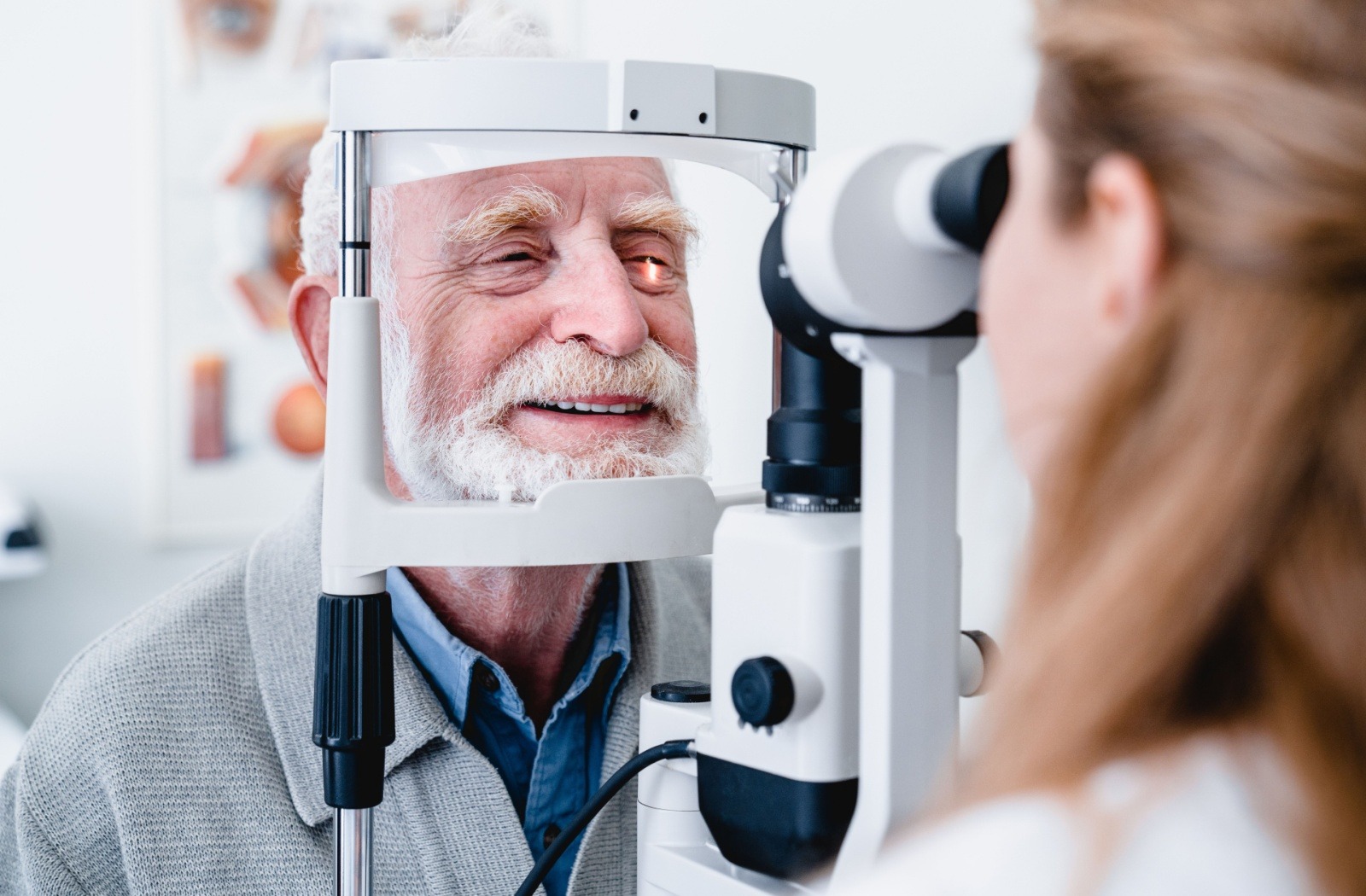
Delivering a diagnosis of dry AMD to your patients brings unique challenges. You want to provide hope alongside honest information about their condition, but traditional management options have focused mainly on slowing progression rather than improving vision.
A comprehensive approach to dry AMD management combines early detection, patient education, lifestyle guidance, and innovative treatment options like MacuMira to help your patients maintain their quality of life and independence. Understanding the complete dry AMD treatment landscape empowers you to provide better care.
What Eye Care Professionals Need to Know About Dry AMD
Dry vs. Wet AMD: Key Differences for Your Practice
Dry AMD affects about 90% of your AMD patients and progresses slowly as the macula thins over time. You’ll notice drusen deposits under the retina during examination, and your patients typically experience gradual central vision changes.
Wet AMD moves much faster and involves abnormal blood vessels that leak fluid into the retina. While less common, wet AMD can lead to rapid vision loss and requires immediate intervention. Your patients with dry AMD face a 10-15% chance of developing the wet form.
Common Signs & Symptoms to Watch For
Early-stage dry AMD often shows no symptoms, making your regular examinations important for detection. As the condition progresses, your patients may report:
- Mild blurriness in central vision
- Trouble seeing in low light
- Straight lines that appear wavy or crooked
- Blurry or blank spots in field of vision
- Colours that seem less bright
Risk Factors That Matter Most
Age remains the strongest risk factor, with most cases appearing after 55. Family history significantly increases risk — patients with affected relatives face up to a 50% higher chance of developing AMD.
Smoking doubles the risk and accelerates progression. Other factors include high blood pressure, obesity, and prolonged sun exposure without eye protection.
The Diagnostic Process: Tools & Techniques for Eye Care Professionals
Complete Eye Exam Protocols
Your comprehensive exam should include visual acuity testing, dilated fundus examination, and careful assessment of the macula. Look for drusen deposits, pigment changes, and any signs of geographic atrophy.
Document the size, number, and location of drusen carefully. Small drusen may indicate normal ageing, while large or numerous drusen suggest AMD progression.
Key Diagnostic Tests You Should Use
Optical coherence tomography (OCT) provides detailed cross-sectional images of retinal layers, helping you detect early changes before symptoms appear. You can measure retinal thickness and track progression over time.
Fundus autofluorescence imaging highlights areas of retinal pigment epithelium dysfunction. This test helps you identify patients at higher risk for progression to advanced stages.
How to Help Patients Use the Amsler Grid at Home
Give your patients an Amsler grid to monitor vision changes between appointments. Show them how to test each eye separately while wearing their reading glasses.
Explain that they should contact you immediately if straight lines appear wavy, bent, or broken. New blank spots or changes in the central area also warrant urgent attention. Learn more about patient education resources that can support your practice.
The 3 Stages of Dry AMD & What to Expect
Early, Intermediate & Late Stage Characteristics
Early-stage AMD shows small to medium drusen with no vision loss. Your patients typically don’t notice symptoms at this stage, highlighting the importance of regular screening.
Intermediate AMD features larger drusen or pigment changes in the retina. Some patients begin experiencing mild central vision problems. Late-stage AMD involves geographic atrophy with significant central vision loss.
Typical Progression Timelines
Progression varies significantly among patients. Some people remain in early stages for decades, while others advance more quickly. About 10-20% of intermediate AMD cases progress to advanced stages within five years.
Monitor your intermediate-stage patients every 6-12 months. Those with large drusen or pigment changes need more frequent follow-ups to catch any rapid progression. Understanding clinical research on progression patterns can inform your monitoring schedules.
Geographic Atrophy: When & How It Develops
Geographic atrophy represents advanced dry AMD where retinal cells die and create blind spots in central vision. This irreversible condition typically develops slowly over several years.
You’ll see well-defined areas of retinal pigment epithelium loss on examination. These areas tend to enlarge gradually, affecting more of your patient’s central vision over time.
Current Treatment & Management Strategies for Your Patients

AREDS2 Supplements: Who Benefits Most
Recommend AREDS2 supplements for patients with intermediate AMD or advanced AMD in one eye. The specific formula includes vitamin C, vitamin E, lutein, zeaxanthin, zinc, and copper.
These supplements can reduce the risk of progression to advanced AMD by about 25%. Patients with early-stage AMD don’t typically benefit from supplementation unless their diet lacks these nutrients.
Lifestyle Recommendations You Can Share
Encourage your patients to stop smoking immediately — it’s the most important modifiable risk factor. A Mediterranean-style diet rich in leafy greens, fish, and colourful vegetables supports eye health.
Regular exercise helps maintain healthy blood flow to the retina. UV protection through quality sunglasses also helps reduce long-term damage from light exposure.
Vision Adaptation Tools & Resources
Connect your patients with low vision specialists when they begin experiencing functional vision loss. Magnifiers, enhanced lighting, and high-contrast materials can help maintain independence.
Modern smartphones and tablets offer excellent accessibility features including voice commands and screen magnification. Many patients benefit from learning these technologies early in their AMD journey. Explore patient success stories to share hope and practical insights with your patients.
MacuMira: A New Treatment Option for Dry AMD
How MacuMira Works in Your Practice
MacuMira uses precisely delivered microcurrent to stimulate retinal pigment epithelium cells, enhancing mitochondrial activity to support cellular energy production.
The device is regulated by the Therapeutic Goods Administration (TGA) and included on the Australian Register of Therapeutic Goods (ARTG), and is regulated by Medsafe in New Zealand, offering a novel treatment option for preserving or improving visual function in patients with dry age-related macular degeneration.
The treatment integrates easily into your existing workflow. Each session takes about 32 minutes, and the non-invasive procedure requires no needles or surgical intervention.
Patient Benefits & Treatment Experience
Your patients experience a comfortable, painless treatment that is indicated to improve visual function. The microcurrent therapy works at the cellular level to support retinal health and function.
Many patients appreciate having an active treatment option rather than just monitoring progression. The treatment schedule fits into most daily routines without disrupting their lifestyle. Learn more about MacuMira therapy and how it transforms patient care.
Training & Support for Your Team
MacuMira provides comprehensive staff training to help your team deliver this dry age-related macular degeneration treatment effectively. You’ll receive ongoing support throughout the lifetime of your device.
The training covers device operation, patient selection, treatment protocols, and how to explain the therapy to your patients. Your team gains confidence in offering this groundbreaking treatment for age-related macular degeneration.
MacuMira represents a significant advancement in dry AMD care, giving you the ability to offer real treatment rather than just hope. With proper diagnosis, monitoring, and access to innovative treatments like MacuMira, you can help your patients maintain their vision and independence for years to come. Consider partnering with MacuMira to expand your treatment options and offer hope to more patients.